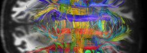
Van Wedeen MD, Harvard Medical School, “Uncovering the Physical and Mathematical Principles of the Heart and the Brain With Diffusion MRI”
Jump to:
Bio
“Uncovering the Physical and Mathematical Principles of the Heart and the Brain With Diffusion MRI “
Van Wedeen is an Associate Professor of Radiology at the Harvard Medical School and the first Thomas J. Brady Endowed Chair in Radiology. Raised in New York City, Dr. Wedeen attended Harvard College with a Westinghouse Scholarship, concentrating in math, earned his MD at Albert Einstein, a residency in internal medicine at Columbia, and then joined Harvard-MGH in 1983 as one of their first fellows in NMR imaging. His work on imaging of motion and flow with tagging and phase contrast led in 1985 to the first MRA, published in Science1, and later, to the first tensor MRI, of cardiac strain, in 1989, and the imaging of myocardial fiber architecture and fiber function2. Building on this approach, Dr Wedeen’s lab for the past 20 years has focused on the mapping brain architecture and connectivity, among their firsts, they presented MRI tractography3 in 1995, developed high angular resolution diffusion MRI4 in 1999-2000, and innovations including DENSE, SMS, and balanced gradients. This program laid foundations for the Connectome Scanner and the Human Connectome Project5, in which Dr. Wedeen was a PI. Recently, this research, using a new analysis of diffusion MRI of the brain, has led Dr. Wedeen’s lab to a new hypothesis about brain architecture. Cerebral axons, they suggest, follow three axes of smooth Cartesian coordinate systems by means of parallel curved sheets – basic mathematical structures called foliations6. If correct, this architecture could provide coordination and authentication of brain structure and connectivity on microscopic and macroscopic scales, leading to new tools of for brain imaging, and potentially clues to brain function, particularly that of the human telencephalon. A focus of Dr. Wedeen’s current research is to further investigate this conjecture, its accuracy and implications, in collaborations of imaging, physics and neuroscience. Holding several patents, Dr. Wedeen is a Fellow of the International Society of Magnetic Resonance in Medicine and Director of Connectomics at the Martinos Imaging Center. Eight of Dr. Wedeen’s students have become professors in radiology and three department chairs.
![]() Click here to view webcast.
Click here to view webcast.
Abstract
“Uncovering the Physical and Mathematical Principles of the Heart and the Brain With Diffusion MRI ”
This year, 2017, honors the centenary of D’Arcy Thompson’s “On Growth and Form”. In it he inaugurates a program to understand living systems using the ideas that revolutionized physics: symmetry and conservation, non-Euclidean geometry, and anticipates broken symmetry, scaling, and emergence, and presumably other ideas yet to be recognized. In this talk, we discuss geometric symmetries in heart and the brain that had been gleaned only with difficulty or in part by classical methods, but which the new methods of diffusion MRI can uncover or make clear.
As an example of symmetry in biological structure, consider the eye and its sphericity. The early development of the eye yields a shape that is fairly round, but the finishing touches are provided by the movement of the eye in its socket, a dynamical system whose function drives structure to greater symmetry. In the heart, morphogenetic programs guide the ventricle to become a cylinder of helical fibers. However, once established, architecture is refined by dynamics. Whereas myocardial deformations vary by 2-3x across the thickness of the wall, cardiocytes converge upon an orientation field given by a toroidal helix, resembling the famed Hopf fibration of SU(2), uniquely to afford almost constant fiber shortenings, of 13±2%. Thus in the heart, universal structure emerges from dynamics.
Recently, fiber architecture of the brain has been noted to show a striking symmetry. Centralized nervous systems arise in bilateral species, and typically have the form of orthogonal grids or ladders coherent with the body axis. The vertebrate is no exception, its nervous system being patterned as an axial checkerboard of regions and connections. Diffusion MRI now suggests this pattern is expressed more extensively and precisely than previously suspected, by an ingenious mechanism. Fiber orientations adhere to coordinate systems by as parallel sheets of crossing fibers, a structure of notable symmetry, called a foliation. We discuss major predictions of this model, and some possible implications for brain mapping and for theories of function. Because we can measure this architecture with dMRI, we may ask how it is distributed among brain systems, and in this way, possibly gain insight into brain function.
![]() Click here to view webcast.
Click here to view webcast.


